2019 / Neuroinformatics / Article
A Web-Based Atlas Combining MRI and Histology of the Squirrel Monkey Brain
Summary
CHIASM combines structural MRI, diffusion MRI, histological stains, and motor-cortex tracing in a common squirrel monkey reference space. The integrated atlas lets researchers compare tissue appearance across modalities and use labeled cortical regions and white matter pathways for anatomical localization.
Study overview
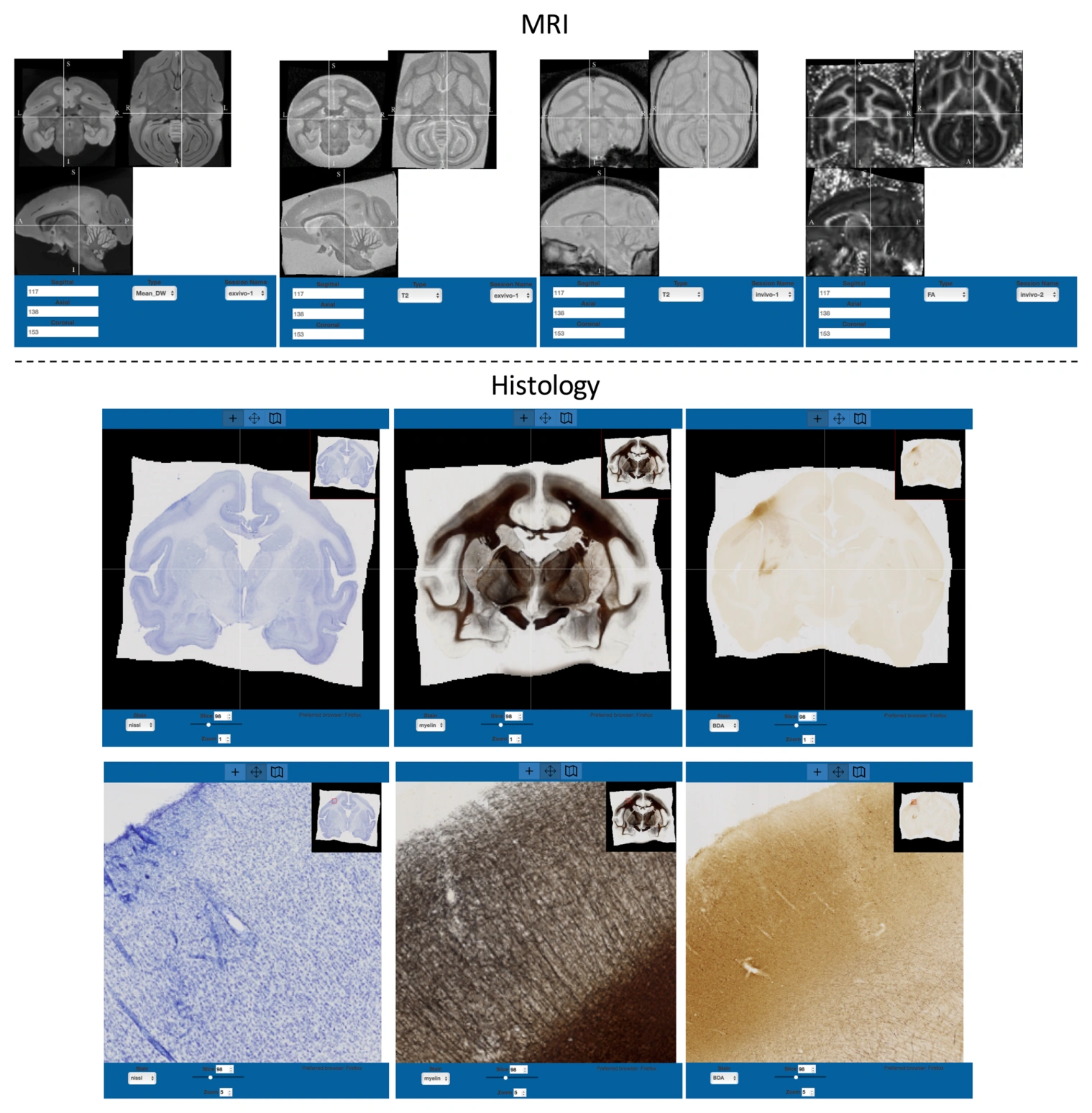
The Combined Histology-MRI Integrated Atlas of the Squirrel Monkey aligns in vivo and ex vivo MRI acquired at 9.4 T with Nissl and myelin histology. A tracer injection in primary motor cortex adds a specific anatomical connectivity reference. The atlas includes diffusion tensor glyphs, 57 tractography-defined white matter labels, and 18 cortical regions identified from cytoarchitecture, accessible through a multimodal web viewer. Bringing these contrasts into one coordinate system supports anatomical interpretation and validation across imaging methods. The reference remains tied to its specimen, registration accuracy, and motor-cortex injection; its tractography labels and histological measurements provide complementary rather than interchangeable evidence.
Main findings
- The atlas combines MRI, Nissl staining, myelin staining, and primary motor-cortex tracer information in one space.
- It includes 57 tractography-defined white matter pathways and 18 histologically defined cortical regions.
- A web-based viewer supports comparisons between user-selected contrasts and resolutions.
Cite this work
Kurt G. Schilling, Yurui Gao, Matthew Christian, Vaibhav Janve, Iwona Stepniewska, Bennett A. Landman, Adam W. Anderson (2019). A Web-Based Atlas Combining MRI and Histology of the Squirrel Monkey Brain. Neuroinformatics. https://doi.org/10.1007/s12021-018-9391-z
This page summarizes the work; consult the original article for the complete methods, results, and qualifications.