2019 / NMR in Biomedicine / Article
Histologically derived fiber response functions for diffusion MRI vary across white matter fibers—An ex vivo validation study in the squirrel monkey brain
Summary
Histology from three squirrel monkey brains shows that diffusion MRI fiber response functions vary across white matter regions. Choosing a different response function changes reconstructed fiber orientations and can alter tractography accuracy.
Study overview
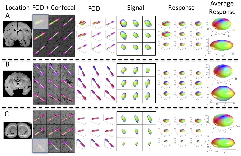
Spherical deconvolution commonly assumes one fiber response function throughout the brain. This study derives reference functions from three-dimensional histology and matched ex vivo diffusion MRI. The functions differ across regions and from conventional estimates, showing that the choice of response function can affect both fiber orientation distributions and reconstructed pathways.
Main findings
- Histologically derived response functions varied across white matter regions.
- Response-function choice changed orientation reconstruction and tractography results.
Cite this work
Kurt G. Schilling, Yurui Gao, Iwona Stepniewska, Vaibhav Janve, Bennett A. Landman, Adam W. Anderson (2019). Histologically derived fiber response functions for diffusion MRI vary across white matter fibers—An ex vivo validation study in the squirrel monkey brain. NMR in Biomedicine. https://doi.org/10.1002/nbm.4090
This page summarizes the work; consult the original article for the complete methods, results, and qualifications.