2026 / Brain Structure and Function / Review
Diffusion tractography outside the brain: the road less travelled
Summary
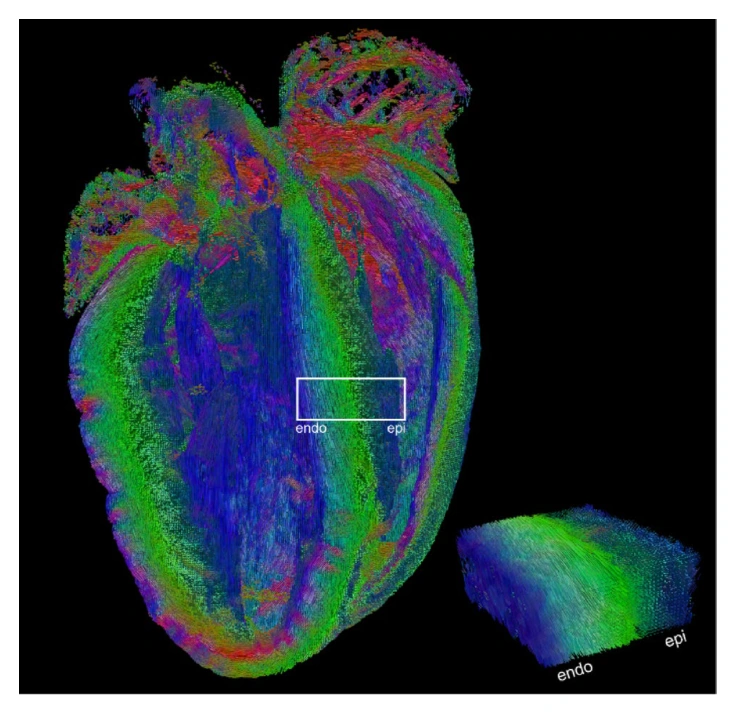
Tractography can probe organized tissue far beyond cerebral white matter. This review surveys applications in the spinal cord, heart, peripheral nerves, brachial plexus, kidney, skeletal muscle, and prostate. It explains the acquisition and modeling adaptations each region requires, together with the anatomical opportunities and unresolved interpretation challenges.
Scope
The organized structure of many tissues creates diffusion patterns that can be investigated with tractography. This review surveys applications outside the brain, including the spinal cord, heart, peripheral nerves, brachial plexus, kidney, skeletal muscle, and prostate. Each application introduces specific acquisition, modeling, and processing requirements, shaped by anatomy and physiology. Motion, susceptibility artifacts, lower anisotropy, and uncertainty about streamline interpretation remain important obstacles. By comparing these settings, the authors describe how diffusion tractography can provide noninvasive information about tissue organization beyond its conventional cerebral applications and identify methodological work needed to strengthen its biomedical use.
Topics covered
- Reviews tractography across seven anatomical regions outside the brain.
- Acquisition and modeling must be adapted to tissue-specific anatomy and physiology.
- Motion, artifacts, and streamline validity remain recurring challenges.
Cite this work
Kurt G. Schilling et al. · Last author: Alexander Leemans (2026). Diffusion tractography outside the brain: the road less travelled. Brain Structure and Function. https://doi.org/10.1007/s00429-025-03062-9
This page summarizes the work; consult the original article for the complete methods, results, and qualifications.