
2025 / Frontiers in Neuroimaging / Article
Leveling up: along-level diffusion tensor imaging in the spinal cord of multiple sclerosis patients
Summary
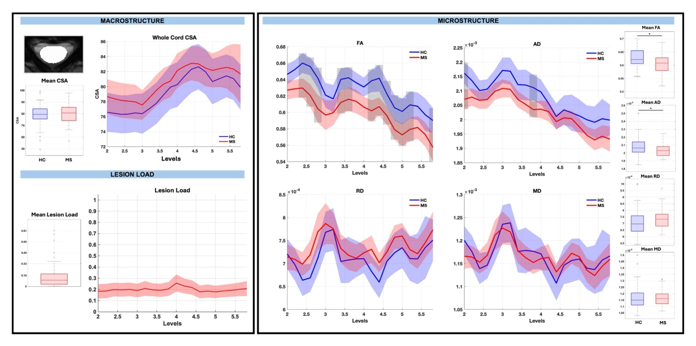
Analyzing spinal cord measurements by cervical level reveals localized changes in relapsing-remitting multiple sclerosis that whole-cord averages can miss. The study combines diffusion and structural measurements within white and gray matter regions. Gray matter atrophy relates to disability, while the measured microstructural changes do not show significant disability associations.
Study overview
The spinal cord has a natural anatomical coordinate system in its cervical levels, providing a way to localize disease-related changes. This study compares people with relapsing-remitting multiple sclerosis and healthy controls using diffusion tensor and macrostructural measurements along the cord. Analyses of white matter tracts and gray matter subdivisions detect spatially restricted differences that are less apparent when measurements are averaged across the whole cord. Gray matter atrophy is associated with clinical disability, whereas microstructural measurements do not show significant correlations with disability in this cohort. The findings support level-specific analysis for characterizing heterogeneous spinal cord pathology.
Main findings
- Level-specific analyses are more sensitive to localized group differences than whole-cord averaging.
- White and gray matter changes vary along levels and across axial regions.
- Gray matter atrophy, but not measured microstructural changes, correlates with disability.
Cite this work
Atlee A. Witt et al. · Last author: Kurt G. Schilling (2025). Leveling up: along-level diffusion tensor imaging in the spinal cord of multiple sclerosis patients. Frontiers in Neuroimaging. https://doi.org/10.3389/fnimg.2025.1599966
This page summarizes the work; consult the original article for the complete methods, results, and qualifications.