
2025 / Magnetic Resonance in Medicine / Review
Considerations and recommendations from the ISMRM Diffusion Study Group for preclinical diffusion MRI : Part 3—Ex vivo imaging: Data processing, comparisons with microscopy, and tractography
Summary
The third ISMRM preclinical diffusion MRI paper addresses ex vivo image processing, comparisons with microscopy, and tractography. It explains how fixed-tissue experiments require choices that differ from in vivo workflows. Community recommendations and unresolved questions guide the design and interpretation of rigorous, reproducible validation studies.
Scope
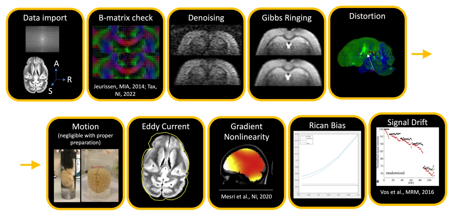
Ex vivo diffusion MRI offers detailed images and opportunities for direct comparison with microscopy, but those advantages introduce distinct processing and interpretation challenges. The final paper in this three-part series covers preprocessing, diffusion modeling, microscopy comparisons, and tractography using ex vivo specimens. It discusses how experimental conditions and tissue preparation influence the resulting measurements and what is needed to connect MRI features to histological evidence. The authors present practical recommendations while identifying areas where firm guidance remains unavailable. The paper supports reproducible validation work and complements the acquisition-focused recommendations in Part 2 of the series.
Topics covered
- Addresses preprocessing and modeling choices specific to ex vivo tissue.
- Discusses comparisons with microscopy and tractography validation.
- Highlights gaps where methodological evidence does not yet support firm guidance.
Cite this work
Kurt G. Schilling et al. · Last author: Ileana O. Jelescu (2025). Considerations and recommendations from the ISMRM Diffusion Study Group for preclinical diffusion MRI : Part 3—Ex vivo imaging: Data processing, comparisons with microscopy, and tractography. Magnetic Resonance in Medicine. https://doi.org/10.1002/mrm.30424
This page summarizes the work; consult the original article for the complete methods, results, and qualifications.