2025 / Magnetic Resonance in Medicine / Review
Considerations and recommendations from the ISMRM diffusion study group for preclinical diffusion MRI : Part 2—Ex vivo imaging: Added value and acquisition
Summary
The second ISMRM preclinical diffusion MRI paper examines the value of ex vivo imaging and how to acquire reliable data. It covers specimen selection, fixation, preparation, and scanning. The recommendations explain both the opportunities for detailed tissue characterization and the experimental differences that complicate comparison with living tissue.
Scope
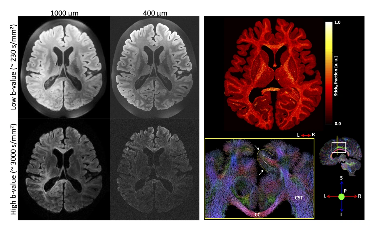
Ex vivo diffusion MRI can achieve high signal-to-noise ratio, fine spatial resolution, and diffusion contrasts that are difficult to obtain in living subjects. It also enables direct comparisons with histology. This review explains those opportunities and the experimental considerations required to use them effectively. Topics include specimen and model selection, tissue fixation, sample preparation, and acquisition protocols. Because ex vivo conditions change tissue properties and measurement requirements, the authors emphasize that in vivo practices cannot simply be transferred unchanged. The recommendations aim to improve reproducibility while identifying remaining gaps in evidence and priorities for future method development.
Topics covered
- Ex vivo imaging enables detailed spatial sampling and advanced diffusion contrasts.
- Fixation and sample preparation materially affect acquisition and interpretation.
- Direct comparison with histology is a major opportunity for validation.
Cite this work
Kurt G. Schilling et al. · Last author: Ileana O. Jelescu (2025). Considerations and recommendations from the ISMRM diffusion study group for preclinical diffusion MRI : Part 2—Ex vivo imaging: Added value and acquisition. Magnetic Resonance in Medicine. https://doi.org/10.1002/mrm.30435
This page summarizes the work; consult the original article for the complete methods, results, and qualifications.