Microstructure
& Connectivity Lab

Publication collection

Spinal Cord

← All publications
2026
NMR in BiomedicineArticle

Standard Model Imaging in the Brain and Spinal Cord of MS Patients: Initial Assessment and Comparison to Diffusion Tensor Imaging

Atlee Witt et al. · Last author: Kurt Schilling

Same-session brain and spinal cord imaging compares standard diffusion tensor measurements with Standard Model Imaging in 34 people with relapsing-remitting multiple sclerosis and 36 controls. Neurite density estimates show sensitivity to pathology in both regions, while the usefulness of other measurements depends on the anatomical setting.

MicrostructureSpinal Cord
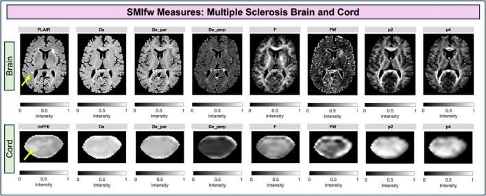
Standard model imaging parameter maps in the brain and spinal cord of a person with relapsing-remitting MS.
2026
Brain Structure and FunctionReview

Diffusion tractography outside the brain: the road less travelled

Kurt G. Schilling et al. · Last author: Alexander Leemans

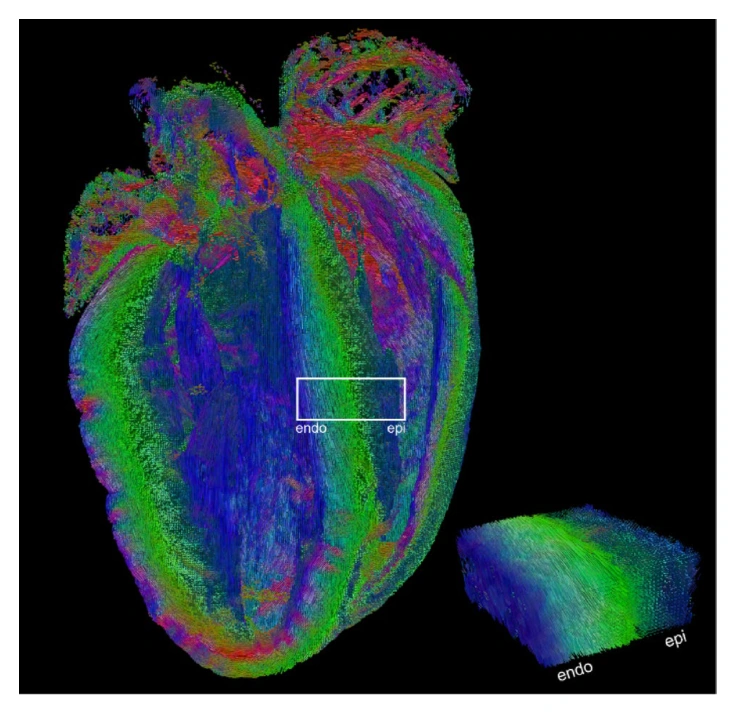
Tractography can probe organized tissue far beyond cerebral white matter. This review surveys applications in the spinal cord, heart, peripheral nerves, brachial plexus, kidney, skeletal muscle, and prostate. It explains the acquisition and modeling adaptations each region requires, together with the anatomical opportunities and unresolved interpretation challenges.

TractographySpinal CordReviews & Consensus
Ex vivo mouse heart tractography shows the changing orientation of cardiomyocytes through the ventricular wall.
2025
Frontiers in NeuroimagingArticle

Leveling up: along-level diffusion tensor imaging in the spinal cord of multiple sclerosis patients

Atlee A. Witt et al. · Last author: Kurt G. Schilling

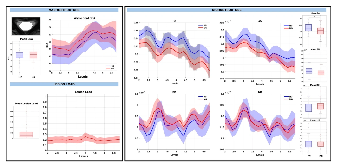
Analyzing spinal cord measurements by cervical level reveals localized changes in relapsing-remitting multiple sclerosis that whole-cord averages can miss. The study combines diffusion and structural measurements within white and gray matter regions. Gray matter atrophy relates to disability, while the measured microstructural changes do not show significant disability associations.

Spinal CordMicrostructure
Whole-cord morphometry, lesion load, and diffusion measurements in healthy controls and people with relapsing-remitting MS.
2025
Imaging NeuroscienceArticle

Characterization of neurite and soma organization in the brain and spinal cord with diffusion MRI

Kurt G. Schilling, Marco Palombo, Atlee A. Witt, Kristin P. O’Grady, Marco Pizzolato, Bennett A. Landman, Seth A. Smith

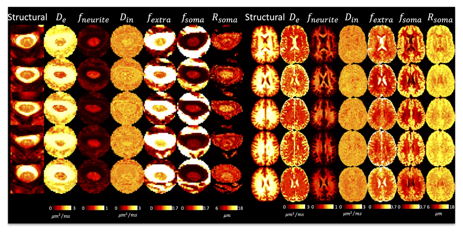
A shared 3T diffusion MRI protocol characterizes brain and spinal cord microstructure using DTI, Standard Model Imaging, and SANDI. The study evaluates image quality, tissue contrast, and repeatability across the neuroaxis. It establishes the feasibility of combined measurements while identifying greater reproducibility and partial volume challenges in the cord.

MicrostructureSpinal Cord
SANDI neurite and soma parameter maps in the brain and cervical spinal cord.
2025
2025 IEEE 22nd International Symposium on Biomedical Imaging (ISBI)Conference paper

Measuring Impact of Super-Resolution on Spinal Cord MRI Scans: Lesion Detection Sensitivity, Variability, and Clinical Impact

Greyson A. Wintergerst, Samuel W. Remedios, Allen T. Newton, Seth A. Smith, Bennett A. Landman, Kurt G. Schilling

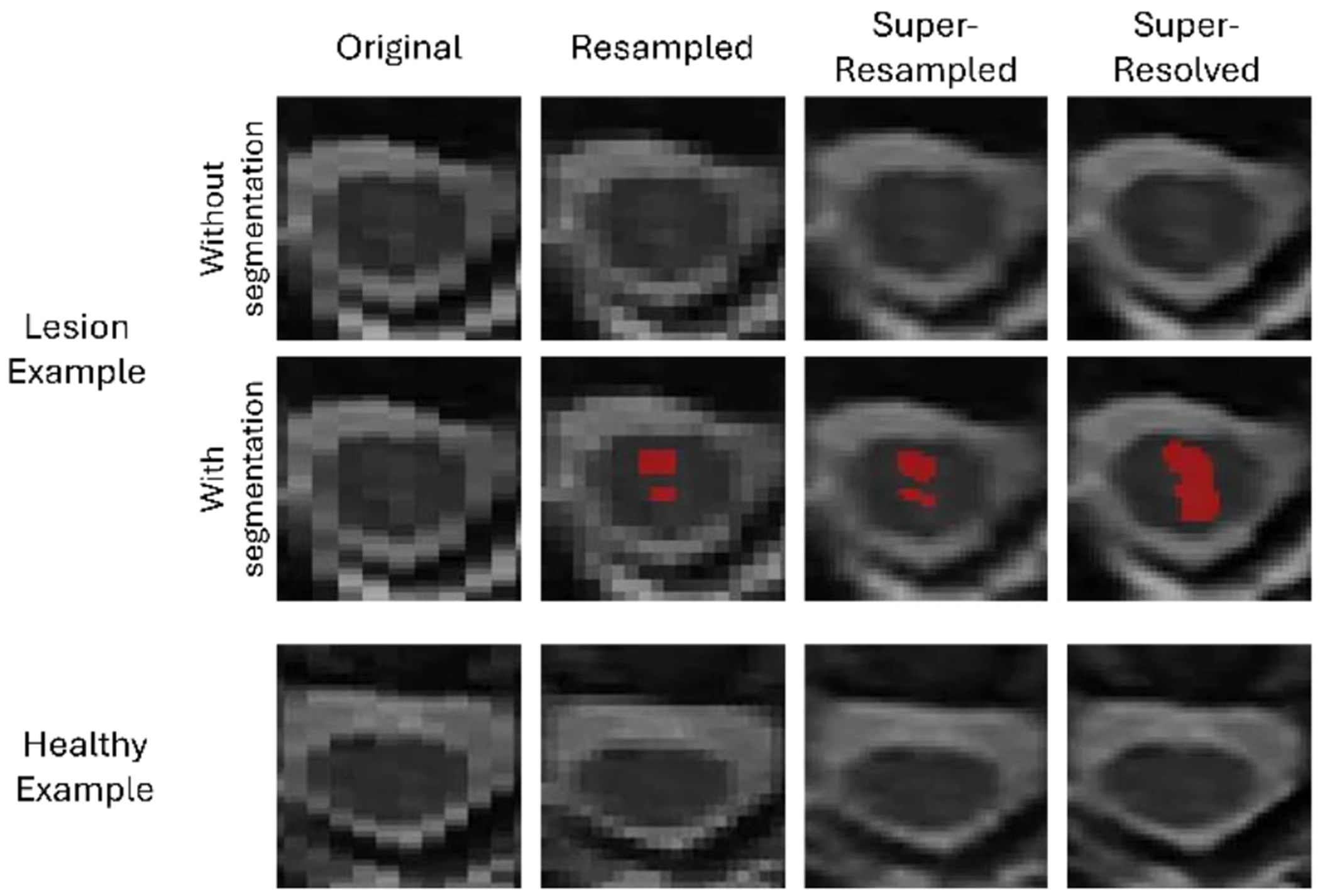
This study tests whether super-resolution and interpolation improve automated lesion detection in spinal cord MRI from 53 people with multiple sclerosis. Higher isotropic resolution increased lesion-segmentation sensitivity, but lesion measures did not show a significant association with disability scores.

Spinal CordImage Processing
Spinal cord image resolution influences the visibility and segmentation of MS lesions.
2024
Magnetic Resonance ImagingArticle

Influence of preprocessing, distortion correction and cardiac triggering on the quality of diffusion MR images of spinal cord

Kurt G. Schilling et al. · Last author: Kristin P. O’Grady

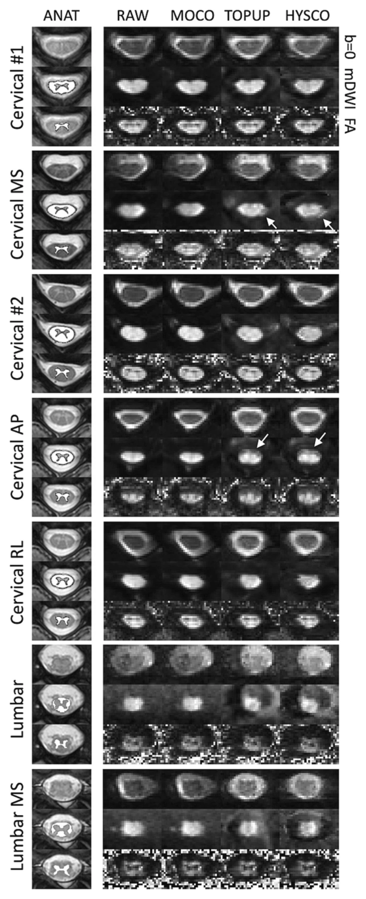
Tests of spinal cord diffusion preprocessing show that better overall image alignment does not necessarily improve geometry or tissue contrast within the cord. Separate experiments found that omitting cardiac triggering shortened cervical acquisitions while preserving similar image quality and diffusion measurements under the conditions studied.

Spinal CordImage Processing
Examples of spinal cord image geometry and contrast after alternative distortion-correction approaches.
2023
NeuroImageArticle

Denoising of diffusion MRI in the cervical spinal cord – effects of denoising strategy and acquisition on intra-cord contrast, signal modeling, and feature conspicuity

Kurt G. Schilling et al. · Last author: Kristin P. O’Grady

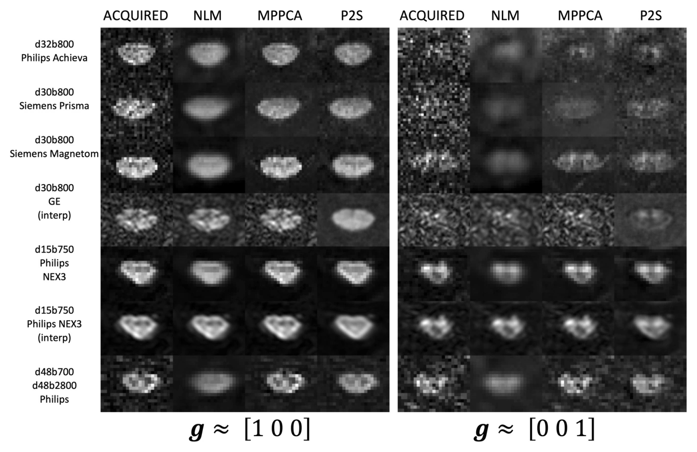
Phantom and human experiments compare three denoising methods for spinal cord diffusion MRI. MPPCA and Patch2Self improved image quality, tissue contrast, and precision even with the small direction counts common in clinical scans, while all tested methods improved visibility of multiple sclerosis lesions in diffusion-weighted images.

Spinal CordImage ProcessingMicrostructure
Spinal cord diffusion images before and after denoising across vendors and acquisition settings.
2022
Magnetic Resonance ImagingArticle

Minimal number of sampling directions for robust measures of the spherical mean diffusion weighted signal: Effects of sampling directions, b-value, signal-to-noise ratio, hardware, and fitting strategy

Kurt G. Schilling, Marco Palombo, Kristin P. O'Grady, Anna J.E. Combes, Adam W. Anderson, Bennett A. Landman, Seth A. Smith

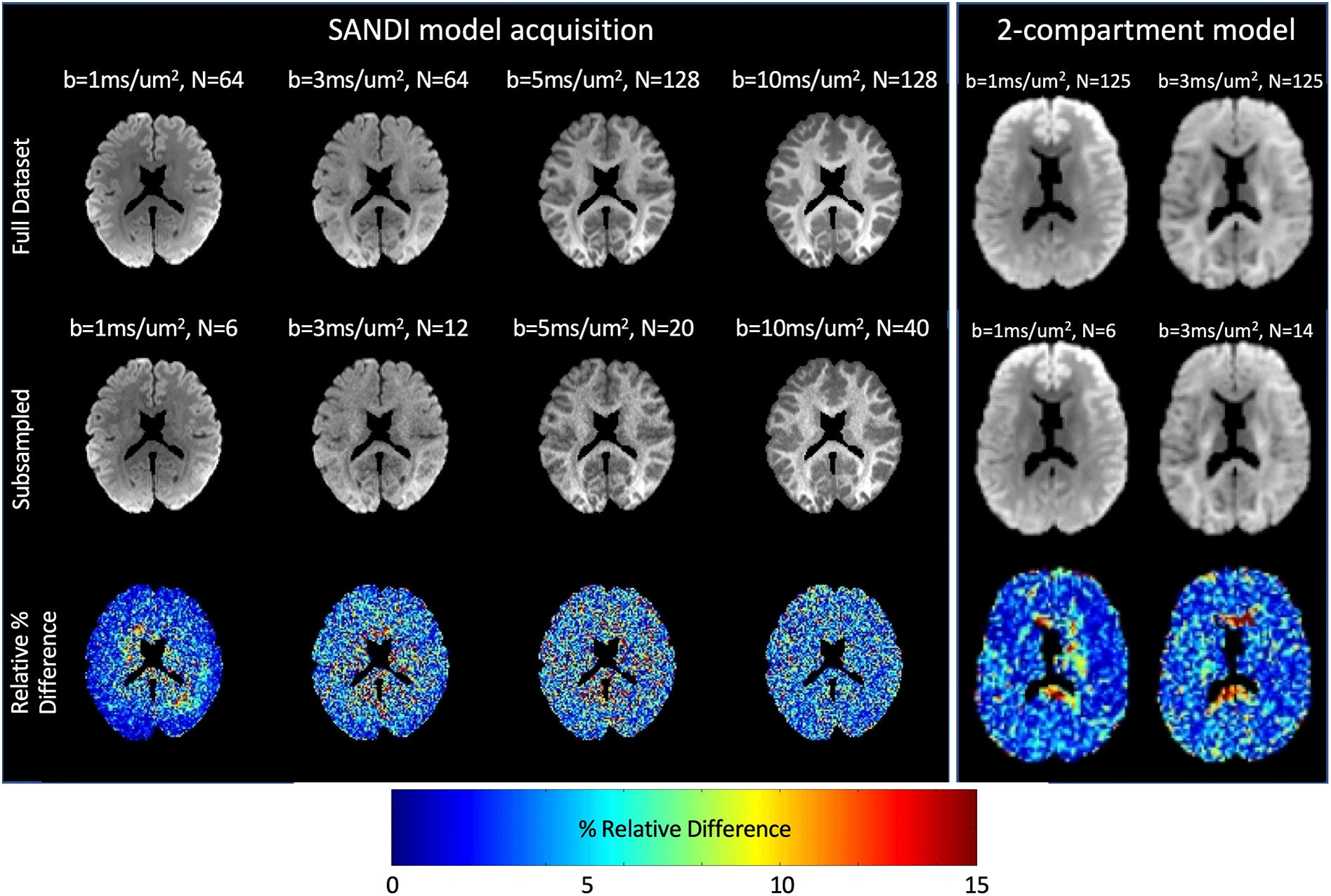
Simulations and brain and spinal cord measurements establish how many diffusion directions are needed to estimate the spherical mean signal reliably. A lookup table links sampling requirements to diffusion weighting, signal-to-noise ratio, and fitting choices.

MicrostructureImage ProcessingSpinal Cord
Brain spherical-mean diffusion signals estimated from full and reduced acquisition schemes.
2019
NeuroImageArticle

Diffusion MRI microstructural models in the cervical spinal cord – Application, normative values, and correlations with histological analysis

Kurt G. Schilling, Samantha By, Haley R. Feiler, Bailey A. Box, Kristin P. O’Grady, Atlee Witt, Bennett A. Landman, Seth A. Smith

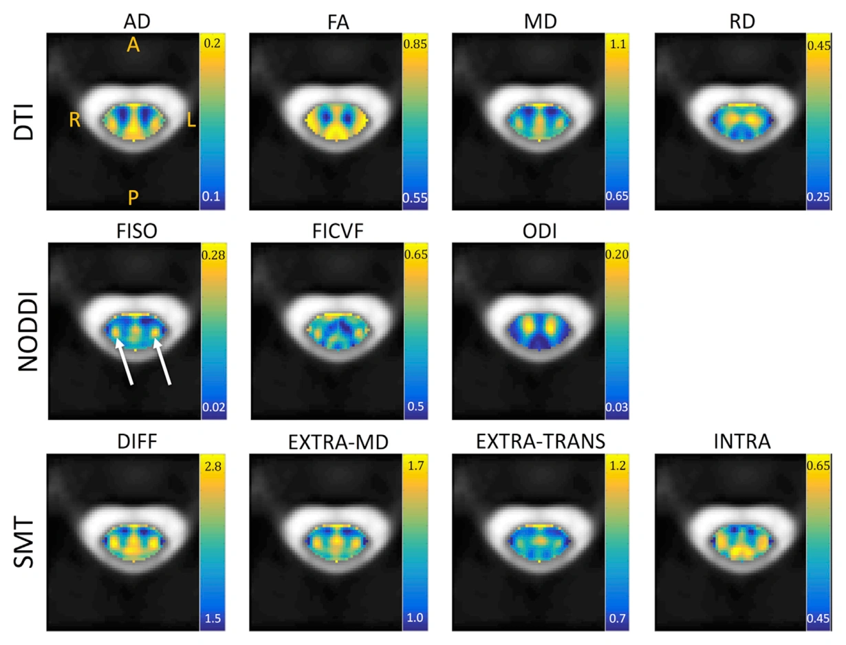
DTI, NODDI, and spherical mean techniques were evaluated in the cervical spinal cords of 21 healthy participants. Comparisons with a histological template showed sensitivity to tissue differences but limited specificity for the biological properties that model parameters are often assumed to measure.

Spinal CordMicrostructure
Population-average DTI, NODDI, and spherical mean technique parameter maps in the cervical spinal cord.